
Multiple Answers, One Dataset
Cryo-TEM imaging can provide quantitative assessment of multiple critical quality attributes of iron nanoparticle formulations all in one study.
Validated methods & data inform batch-to-batch reproducibility including scale-up, comparability of biosimilars & generics, and stability studies.


Cryo-TEM Characterization of Iron Nanoparticles
Cryo-TEM imaging & automated image analysis tools can assess iron nanoparticle CQAs:
- Aggregation
- Morphology
- Size of particles, including dense cores

Nanoparticles We Can Image with Cryo or NS TEM


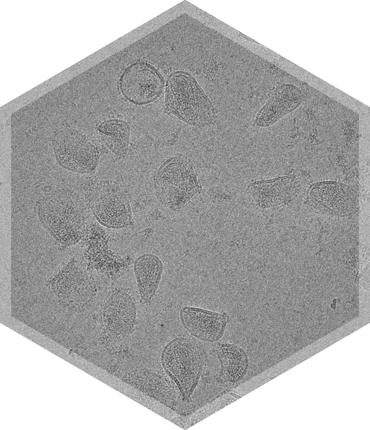


Comprehensive Characterization of Iron Nanoparticles with Cryo-TEM Imaging
Anemia and iron deficiency are commonly observed in the clinic, especially under certain conditions such as pregnancy, chemotherapy, and chronic kidney disease. The delivery of intravenous or intramuscular iron supplements to a patient must be carefully controlled to avoid toxic side effects. Beginning in the 1940s, it was demonstrated that complexes of carbohydrates and iron oxyhydroxide allow for the rapid but safe delivery of iron. Several different formulations have since been developed and approved for clinical use. These formulations all form nanoparticles that vary in the composition of their carbohydrate shell and the size and stability of their iron cores. The exact physicochemical characteristics of these nanoparticles can affect their pharmacokinetic profile.
Cryo Transmission Electron Microscopy (cryo-TEM) allows for the assessment of several important features of iron nanoparticle formulations. The size of the entire nanoparticle, as defined by the carbohydrate shell, can be assessed directly from cryo-TEM images. The increased electron scattering of iron relative to carbohydrates causes regions of varying density to show up within the nanoparticle and can potentially allow for estimation of iron core size within these nanoparticles. Using a proprietary in-house developed machine learning algorithm, NIS can potentially size both particles and dense cores in your iron nanoparticle formulation. The aggregation, morphology, and size of particles can all be assessed in cryo-TEM images.

.png)
Frequently Asked Questions

Is your particle sizing analysis able to size my iron nanoparticles and their dense cores?

How many particles do you count, or images do you analyze per sample or grid?


