
Precise Characterization of AAV
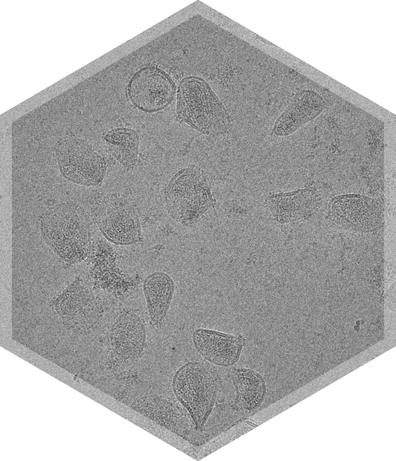
Cryo-TEM imaging paired with proprietary automated analysis tools provide reliable, reproducible, rapid insights into your AAV formulations, including the empty-to-full capsid ratio, aggregation, the presence of contaminants, the presence of unencapsulated DNA, capsid integrity, and more.


Faster, More Informed Decisions About Your AAV Formulations
Cryo-TEM imaging & automated analysis tools can assess:
- Fully automated full/partial/empty capsid ratio analysis, with a variety of payload sizes
- Morphology, including broken capsids
- Aggregation levels
- Sample integrity & impurities, including free DNA
- 3D structure reconstructions


Characterize Adeno-Associated Virus (AAV) with Automated Classification
Adeno-associated virus (AAV) is a well-established platform for gene delivery. It is crucial to ensure that AAV formulations are free from impurities that can compromise the efficacy, safety, and immunogenicity of the product. One such impurity is the presence of empty capsids or capsids with incomplete DNA payloads.
Cryo-TEM imaging combined with quantitative image analysis tools can be used to directly assess the composition of AAV formulations. Automated particle classification analysis of cryo-TEM images, performed with NIS's proprietary machine learning algorithm, enables precise characterization of full and empty capsids. Simultaneously, it is possible to visualize integrity, impurities, and the level of aggregation. Cryo-TEM is the ultimate tool to evaluate combined CQAs; for example, it can be used to assess whether it is empty or full capsids that are more prone to aggregation.
Automated image analysis algorithms allow for reproducible, reliable full/partial/empty capsid ratio data across a variety of payload sizes, which stringly aligns with AUC data. Download our Technical Note to learn more about our automated capsid analysis.

Furthermore, cryo-TEM imaging can be used to obtain 2D class averages and create high resolution 3D reconstructions of AAV particles. This can help identify potential post-translational modifications on AAV capsids, decipher the structural impact of mutations on capsid structure, and help understand the molecular basis of receptor bound AAV or epitope mapping.

Further, we are able perform negative stain TEM imaging of AAV particles. The sample is stained with a heavy stain, such as uranyl formate, and results in high contrast images suitable for detecting impurities and the integrity of particles, including discreet capsomers.


Frequently Asked Questions

How many particles do you count, or images do you analyze per sample or grid?

Can I see an example report for your automated capsid ratio analysis?
Is cryo-TEM analysis of AAV capsid ratios repeatable?
How does cryo-TEM compare with AUC for AAV full/empty/partial capsid ratio analysis?


