
Per-Particle Visualization of Adenovirus
Cryo-TEM imaging & analysis of adenovirus formulations provide unique insights for development & quality control.
Validated methods & data can help assess the impact of factors like addition of excipients, scale-up procedures, and storage conditions (time, temperature, buffer, etc.) on the efficacy, stability, and overall success of adenovirus formulations.


Adenovirus Characterization with Cryo-TEM
Rapid, reproduceable assessment of multiple adenovirus CQA’s with one cryo-TEM dataset:
- Size
- Morphology
- Aggregation status
- Purity
- Structural integrity
- High resolution 3D structures


Visualize Adenovirus formulations on a per-particle level, from cryo-TEM images to 3D structure determination.
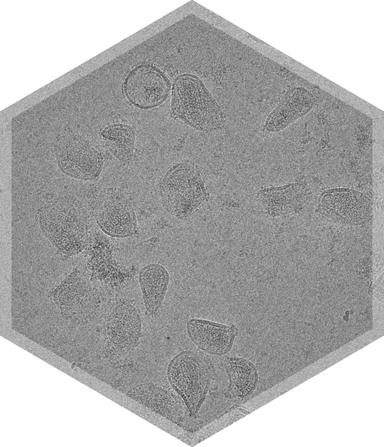
Cryo-TEM imaging and analysis is a powerful tool for characterizing Adenovirus (AdV), and can be used to study morphological characteristics as well as provide high resolution structures.
Cryo-TEM can help assess size, morphology, aggregation status, purity, and structural integrity of adenovirus formulations. This characterization is useful in development as well as quality control and stability assessments, as factors including the addition of excipients, scale-up procedures, and storage conditions (time, temperature, buffer, etc.) can significantly impact the efficacy, stability, and overall success of adenovirus formulations.
Characterizing adenovirus with cryo-TEM has gained traction in recent years, particularly due to the development of AdV based COVID-19 vaccines like those from AstraZeneca and Janssen.
Particle classification analysis of cryo-TEM images can be used to categorize and quantify morphological features in and between formulations, such as revealing surface decorations and proving proof of concept of modular decoration of adenovirus capsid surfaces with antigens.
High resolution cryo-TEM imaging and single particle analysis can also provide detailed 2D classification and the full 3D structure reconstruction. A 3D structure can provide information on for example antibody binding sites or epitope mapping, if and how mutations affect the capsid structure, receptor binding sites, and possibly post-translational modifications.
See an example of adenovirus characterization done by NIS in the paper “Modular capsid decoration boosts adenovirus vaccine-induced humoral immunity against SARS-CoV-2” by Dicks, Matthew D.J. et al. (https://doi.org/10.1016/j.ymthe.2022.08.002)

.png)
Frequently Asked Questions

How many particles do you count, or images do you analyze per sample or grid?


