
Per-Particle Analysis of Exosomes & Extracellular Vesicles
Cryo-TEM can provide detailed insights into exosome & extracellular formulations in their near native state, with only ~50μl of sample. Direct visaulization of exosomes and EVs allows insights into complex formulations to inform lot comparisons, batch-to-batch scale up, and stability studies.
Quantitative and qualitative data, along with method validated studies, provides a deeper understanding of EV and exosome formulations.


Samples Related to Extracellular Vesicles & Exosomes
Characterize Extracellular Vesicles & Exosomes with Cryo-TEM Direct Imaging
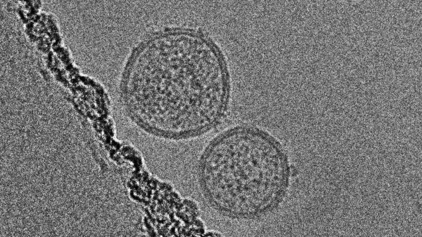
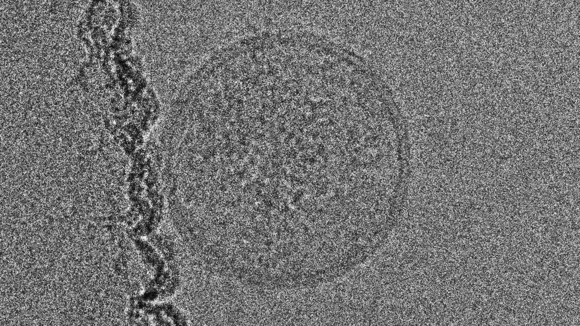
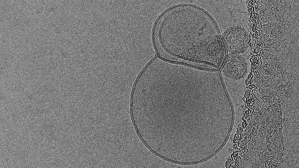
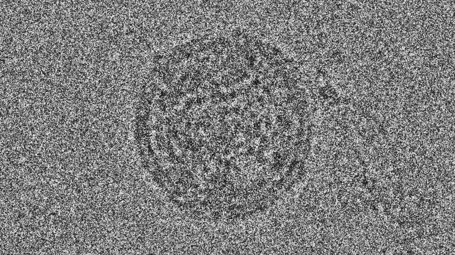
Extracellular vesicles (EVs), including exosomes, have been discovered to play an important role in intercellular communication between tissues in the body. Numerous innovative approaches are being developed to deliver therapies using these biologically-derived structures. EVs/exosomes are derived from numerous sources, including body fluids (e.g., blood plasma, amniotic fluid, milk, etc) and cell culture media (including mesenchymal stem cells and fibroblasts). Endogenous EVs/exosomes carry nucleic acids, proteins, and lipids, which can all potentially affect target cells. Additionally, EVs/exosomes can be loaded with therapeutic agents to modify or enhance their mode of action.
Depending on how source materials are processed, the composition of the final drug substance can be quite complex. Characterization of EV/exosome products can therefore be challenging. Size distribution, lamellarity, cargo encapsulation, and stability are all critical attributes of EVs/exosomes that can be assessed using cryo-TEM direct imaging.
EVs/exosomes can range in size from 50 nanometers to hundreds of nanometers, depending on their type, source, and cargo. They can also vary in structure, internal cargo, and surface decoration. Unlike any other technique, cryo-TEM allows for direct visualization of each of these attributes. Size distribution analysis and particle classification studies can be used to compare lots and study stability. Immunogold labeling can be performed to confirm the presence of proteins on the surface of EVs/exosomes.
Frequently Asked Questions

How many particles do you count, or images do you analyze per sample or grid?


