
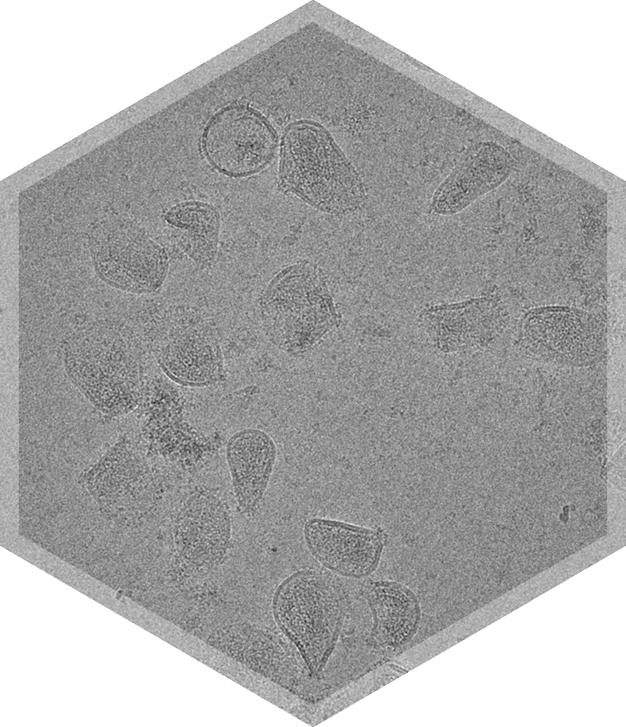
Qualitative Morphology Assessment with Cryo-TEM
Cryo-TEM provides per-particle insights into morphological characteristics of nanoparticles:
- Particle shape & circularity
- Quantitative particle classification, including multivesicular or multicompartmental particles
- Irregular particles & unexpected results
- Integrity, impurities
- Level of aggregation
- Formulation uniformity
.png)

.png)

Samples We Can Perform Morphology Analysis On


Complete Morphology Assessment with Cryo-TEM
Direct determination of particle morphology is a critical aspect of nanoparticle characterization. Typical analyses include particle size distribution, shape, and overall appearance of particles in a sample, and can include quantitative analysis of circularity.
All of these measurements can be obtained in one cryo-TEM or negative stain imaging study, with a small amount of sample.


Frequently Asked Questions

Can I see an example report showing your morphology & lamellarity analysis?

How many particles do you count, or images do you analyze per sample or grid?

