
.png)
Revealing the 3D Structure of Nanoparticles with Cryo-Electron Tomography
If you work with complex nanoparticles, you’ve likely run into a familiar frustration: two-dimensional (2D) cryo-TEM images show you something interesting, but you can’t quite tell what’s going on inside the particles or why some particles in some formulations look different from others.
Standard cryo-TEM provides a 2D projection of a 3D object. That's powerful for many characterization tasks, including sizing, fraction counting, and payload assessment. But when the question depends on understanding 3D morphology, 2D projections can fall short. Examples where 3D analysis becomes important include determination of particle volume for non-spherical particles, examination of internal structures, and identification of surface modifications of particles.
That's where cryo-electron tomography (cryo-ET) comes in.
What Is Cryo-Electron Tomography?
Cryo-ET is a technique for generating true three-dimensional reconstructions of individual nanoparticles, preserved in their native hydrated state. Think of it like a CT scan in medicine: instead of a single X-ray image, many images taken from different angles are combined into a full 3D volume that can be explored slice by slice.
If you’re familiar with single particle analysis (SPA), the concept of building a 3D structure from 2D cryo-EM images may already be intuitive. But SPA relies on imaging thousands of copies of an identical particle in different orientations and computationally combining them. This is an approach that works beautifully for proteins, but breaks down for complex nanoparticles like LNPs, lentiviral vectors, or baculoviruses, where every particle is structurally unique.
Cryo-ET takes a different approach. Instead of relying on finding examples of the particle in many different orientations across many images, it collects a series of 2D images of the same field of particles as the field is tilted from -60 to +60 degrees in increments of 2 or 3 degrees (Figure 1).
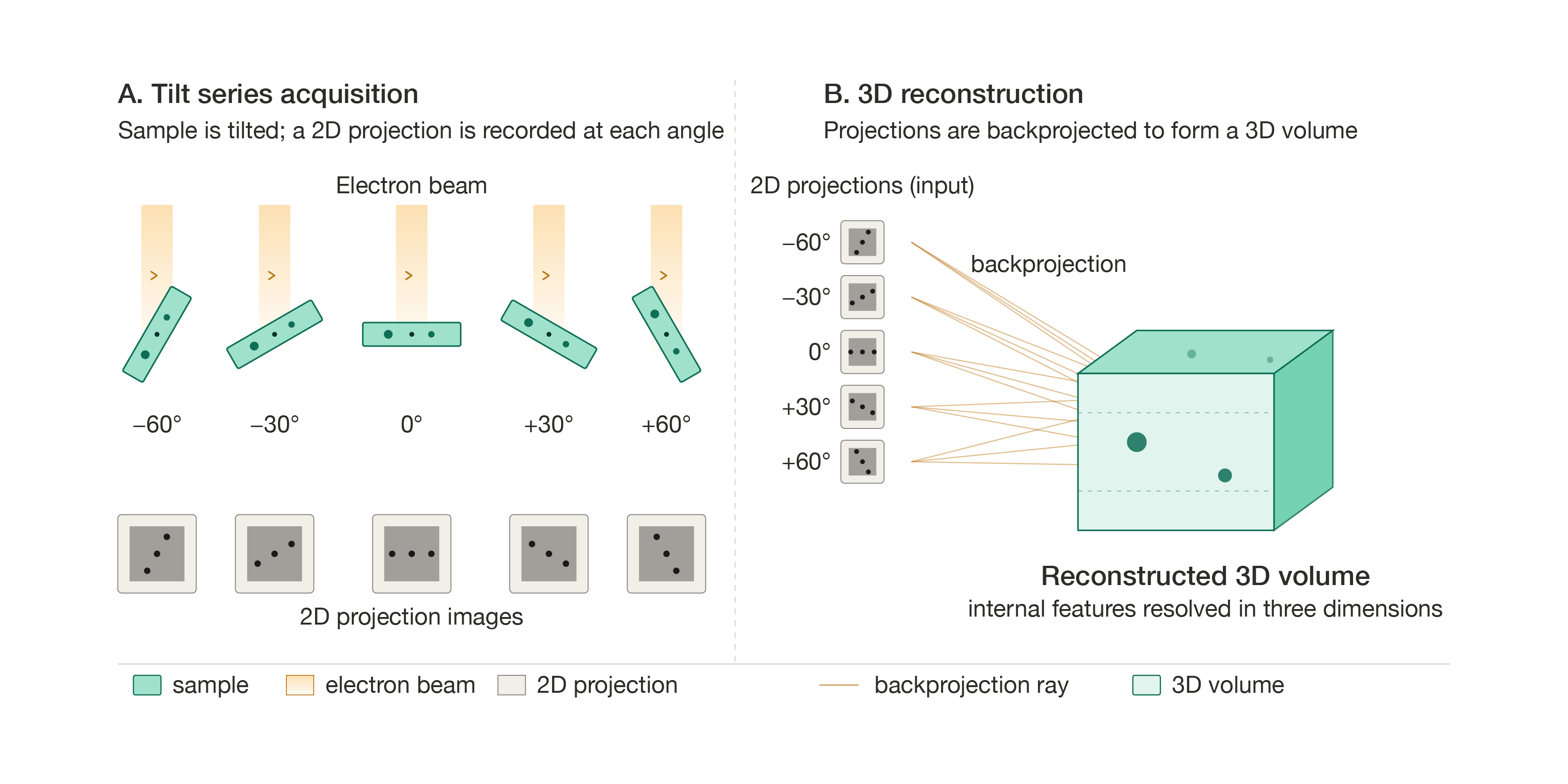
Those images are then aligned and reconstructed into a 3D volume called a tomogram. The result is a complete, sliceable 3D dataset that captures each particle’s individual architecture. These volumes have a resolution of 2 to 3 nm in the XY plane, and slightly worse resolution in the Z plane due to the inability to collect a tilt series extending from -90 to +90 degrees. In practice, the Z-resolution is still sufficient for the structures most relevant to nanoparticle characterization such as lipid bilayers, internal compartments, and surface features.
How the Process Works
The sample preparation for cryo-ET is the same as for standard cryo-TEM. A small volume of sample (3 microliters) is applied to a grid, blotted to a thin film, and rapidly plunge-frozen in liquid ethane using an automated vitrification robot. This preserves particles in amorphous (vitreous) ice, maintaining their native structure.
Once the grid is loaded into the electron microscope, a suitable area is identified, and the tilt series is collected. After acquisition, the individual tilt images are computationally aligned to correct for any sample movement during collection, and then a reconstruction algorithm (usually a method called weighted back projection) generates the final 3D volume.
The tomogram can then be explored slice by slice, rendered as a 3D volume, or further analyzed through object segmentation, where structures of interest are traced and measured to extract quantitative data like volume, surface area, and spatial relationships between particle features.
Case Study 1: Lentivirus
Key finding: Cryo-ET quantified that 71% of lentiviral particles contained an intact capsid, which was impossible to determine from 2D projections alone. The internal features of lentiviral particles can be challenging to interpret in 2D cryo-TEM images. In this particular sample, the particles are dense due to a variable environment inside the mature virion: the capsid is enclosed within a pleomorphic envelope containing viral envelope glycoproteins, matrix proteins, and a condensed core of ribonucleoprotein complexes, all of which create significant signal-to-noise ratio challenges in standard 2D cryoEM. In addition, the particles are surrounded by lipid and protein debris throughout the sample. These features can make it very difficult to confirm whether an intact capsid is present inside the envelope. Since capsid presence is known to vary in lentiviral preparations and is a critical attribute of lentiviral particles, this is an important characterization question.
Cryo-ET provided a much clearer depiction of the sample. In the tomographic slices, the conical capsid structures were clearly visible within individual viral particles (see Figure 2). Spike proteins on the particle surface were also resolved with high clarity.
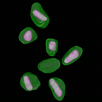
Across 17 tomograms and 104 viral particles, the analysis showed that 71% of particles contained a clearly identifiable capsid. That kind of quantitative assessment of an internal structural feature simply isn't possible from 2D projections of these complex, heterogeneous particles.
Case Study 2: Protein-Modified LNPs (Unsuccessful Conjugation)
Key finding: Cryo-ET confirmed that surface conjugation failed and internal complexity suggested by 2D imaging is resolved by tomography. An LNP formulation was designed with a surface protein for targeted delivery. In standard 2D cryo-TEM images, the sample contained abundant free protein throughout the field, making it impossible to determine whether any protein was specifically associated with the particle surfaces. Cryo-ET resolved this question: the protein was randomly distributed through the sample and not localized to particle surfaces. Across multiple tomograms, surface conjugation appeared to have been unsuccessful.
The tomograms also revealed complex internal morphologies, including blebs within blebs and particles nested inside other particles, features that would be very hard to study in 2D (see Figure 3).
Case Study 3: Antibody-Conjugated LNPs (Successful, but Hidden)
Key finding: In contrast to Case Study 2, conjugation had succeeded but the antibody corona was invisible in 2D because of air-water interface migration. Without tomography, a well-labeled formulation would have been incorrectly abandoned. A second LNP formulation was conjugated with antibodies for targeted delivery. The 2D cryo-TEM images were ambiguous: the sample was clean, with faint specks visible near some particle surfaces, but most particles appeared to have little or no labeling.
Cryo-ET told a very different story. In the interior of the vitreous ice film, a few particles had visible antibodies. But at the air-water interface (the very top and bottom surfaces of the ice film), nearly every particle was surrounded by a dense corona of protein. The antibodies had migrated to the air-water interface during sample preparation, a well-known phenomenon in cryo-EM. Viewed from above in a standard 2D projection, this corona is nearly invisible because the protein sits at the poles of the particle, not around its equator.
The 3D segmentation confirmed that the particles were, in fact, robustly labeled (see Figure 4). Without tomography, the formulation might have been incorrectly judged as poorly conjugated.
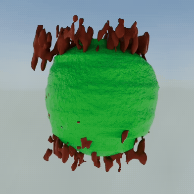
Case Study 4: Identifying an Air-Water Interface-Sensitive Subpopulation
Key finding: Cryo-ET identified a distinct subpopulation of interface-sensitive particles and reveals apparent mRNA distribution within LNPs. A third LNP formulation contained faint, round structures in the 2D images that were difficult to characterize. Cryo-ET revealed two distinct populations. The first consisted of typical LNP particles with lipid bilayers and internal bleb structures. The second was a subpopulation of particles that were disintegrating at the air-water interface, appearing as hemispheres in 3D. These particles consistently lacked a lipid bilayer and appeared to lack blebs, suggesting that their surface composition made them vulnerable to the interface.
The tomograms also resolved fine structural details within the lipid compartments of intact LNPs. Dense thread-like structures running parallel along the subsurface of particles were observed. These structures were 2.5 nm in diameter on average with 8.2 nm spacing between threads. These are consistent with RNA-lipid interaction patterns that have been described in the published literature, and their presence and distribution varied from formulation to formulation.
When to Consider Cryo-ET
Cryo-ET is not a replacement for standard cryo-TEM imaging. It is lower throughput, and the data analysis requires more effort; however, for certain questions, it is the only way to get a definitive answer.
Consider cryo-ET when you need to:
- Distinguish particle interiors from exteriors
- Confirm the presence or absence of internal structures (such as capsids in viral vectors)
- Verify whether a surface modification has been successful
- Characterize complex or unusual particle morphologies
- Understand subpopulations in heterogeneous samples
- Extract quantitative 3D measurements like volume fractions and structural dimensions
Even a quick look at a couple of tomograms can sometimes reveal critical information about your sample. And for more in-depth studies, collecting dozens of tomograms in a single microscope session enables the kind of population-level 3D analysis shown in the LNP examples above.
A Note on Subtomogram Averaging
For samples where a consistent, repeating structural element is present, such as a spike protein displayed on the surface of a viral vector or LNP, there is the possibility of combining cryo-ET with single particle averaging approaches. This technique, known as subtomogram averaging, can potentially yield high-resolution 3D structures of those repeating elements and even resolve conformational heterogeneity within the population. For example, the spike proteins clearly resolved on the surface of lentiviral particles in Case Study 1 would be prime candidates for subtomogram averaging, potentially yielding near-atomic resolution structures of those surface antigens in the context of the intact particle.
Interested in cryo-ET for your viral vector program? Reach out at info@nanoimagingservices.com.
About NIS
NanoImaging Services is a contract research organization with years of experience in cryo-electron microscopy. With facilities in San Diego (NIS-West) and Boston (NIS-East), and protein production capabilities through Proteos in Kalamazoo, NIS provides an integrated path from gene to structure. NIS is the only cGMP-compliant cryo-TEM lab in North America, supporting both research and regulatory submissions.
To discuss a cryo-ET project or learn more about our nanoparticle characterization capabilities, contact us at info@nanoimagingservices.com.
Welcome to Finsweet's accessible modal component for Webflow Libraries. This modal uses Webflow Interactions to open and close. It is accessible through custom attributes and custom JavaScript added in the embed block of the component. If you're interested in how this is built, check out the Attributes documentation page for this modal component.
Infographic Available for Download
Cryo-ET delivers true 3D reconstructions of nanoparticles. See how NIS uses cryo-ET to characterize LNPs, lentiviral vectors, and surface modifications.


