Analyses

Epitope Mapping

Scientific rendering of 3D protein structure, structural biology | Nano Imaging
NIS Molecule
NIS Protein Structure

Unraveling the Molecular Basis of Antibody-Antigen Complexes with Cryo-EM and Negative Stain TEM Imaging

To develop antibody therapeutics and vaccines, researchers need to thoroughly understand how antibodies interact with their target antigens. Epitope mapping, a process that identifies the precise region where antibodies bind to antigens, is essential for unraveling immune responses and designing targeted therapeutic antibodies. By deeply understanding the structure of the epitope, we can uncover atomic interactions and conformational changes, and their impact on the mechanism of action (MOA) of these therapeutic agents.

Cryo-EM: A Revolution in Structural Biology

In structural biology, cryo-electron microscopy (cryo-EM) has emerged as a powerful tool, providing unparalleled resolution and structural details for epitope mapping. By combining cryo-EM imaging with single particle analysis workflows, scientists can confidently solve structures, contributing to our understanding of complex biological systems.

Negative Stain TEM: A Vital Complementary Imaging Technique

Negative stain transmission electron microscopy (TEM) workflows offer additional analysis of antibody antigen complexes for epitope mapping. This technique involves staining the sample with heavy metals to enhance contrast for visual analysis. However, negative stain TEM provides only nanometer-range resolution, which is lower than typical cryo-EM structures. Additionally, the heavy metal stain can often introduce structural artifacts by dehydrating the sample. Despite these limitations, negative stain TEM still plays a vital role in validating the integrity of antigen-antibody complexes and identifying the specific region of the antigen recognized by antibodies. Moreover, it often yields faster results than cryo-EM imaging.

Example workflow of negative stain TEM imaging of IgM particles, with 2D class averages, and focused classification showing an improved IgM average from selective masking (red circles) of sub-classification of particles | epitope mapping by NanoImaging Services
Example workflow of negative stain TEM imaging of IgM particles, with 2D class averages, and focused classification showing an improved IgM average from selective masking (red circles) of sub-classification of particles.

Integrative Imaging: A Comprehensive Look at Antibody-Antigen Interactions

By combining 2D classifications gained through negative stain TEM imaging and high resolution 3D models and maps from cryo-EM imaging on ThermoFisher Krios microscopes, researchers gain a wealth of information about their antibody-antigen complexes. This multi-faceted approach provides a comprehensive understanding of the intricate structure of these critical biological interactions.

Learn More About Antibody-Antigen Complexes:

  • Antibody-Antigen Complexes
  • Antibody-Antigen Complexes

No items found.

Frequently Asked Questions

Explore how our experts can help you.

NIS Molecule

What are the deliverables for NIS's rapid Epitope Mapping service?

Menu Hex
what-are-the-deliverables-for-niss-rapid-epitope-mapping-service

The rapid Epitope Mapping service deliverables include a 3D reconstruction to a resolution at the epitope/paratope interface suitable for tracing the chains and assigning 75% of the side chains. This could include a docked antigen and FAB molecules, no refinement. This is provided within two weeks of sample receipt.

Why should I use cryo-EM for Epitope Mapping instead of Hydrogen-Deuterium Exchange (HDX)?

Menu Hex
why-should-i-use-cryo-em-for-epitope-mapping-instead-of-hydrogen-deuterium-exchange-hdx

HDX can offer faster initial results compared to cryo-EM, particularly when preliminary work involving vitrification conditions and grid screening needs to be done. However, once these conditions are established, the turnaround times for both techniques can be similar. NIS is able to provide results in 2 weeks or less from sample receipt.

It's important to note that HDX provides "low-resolution" details, primarily revealing the general area of interactions at a peptide level. In contrast, cryo-EM has the capability to provide specific atomic interactions. In certain cases, HDX profiles are not distinct enough to differentiate between antibodies targeting similar epitopes, whereas cryo-EM is able to visualize the different binding orientations of antibody-antigen complexes.

Learn more about the differences between techniques in our blog here, or webinar with CSO, Dr. Giovanna Scapin here.

Why should I use cryo-EM for Epitope Mapping instead of X-ray Crystallography (XRC)?

Menu Hex
why-should-i-use-cryo-em-for-epitope-mapping-instead-of-x-ray-crystallography-xrc

X-Ray Crystallography has traditionally been used for epitope mapping. XRC can give 3 dimensional, atomic level interactions, and provide detailed information about the binding sites.

However, not all antibody-antigen complexes are able to crystallize. Cryo-EM is able to map complexes that will not crystallize, and can be a better technique for samples that are too large, flexible, or dynamic for XRC. With NIS’s two week turnaround time, cryo-EM can provide atomic resolution details rapidly.

Learn more about the differences between techniques in our blog here, or webinar with CSO, Dr. Giovanna Scapin here.

What are obstacles you see in epitope mapping with cryo-EM?

Menu Hex
what-are-obstacles-you-see-in-epitope-mapping-with-cryo-em

NIS sees preferred orientation in almost 30% of our vitrified samples, however, over the years have developed a number of strategies including the use of detergents, or changing vitrification methods. To read more about our strategies for overcoming preferred orientation, read our blog here.

If the sample size is under 80 kDa, it will be too small to visualize well. There are strategies for breathing more mass, such as binding multiple antibodies to one antigen, however, small complexes are often not good candidates for cryo-EM.

If your sample concentration is under micromolar, we tend to see dissociation so we ask for 0.5-5 mg/mL concentration. However, if your sample is outside of these parameters, we may be able to use a higher concentration of one component of the sample, or use the SPT Labtech Chameleon for vitrification which allows a high protein concentration than other vitrification methods.

What sample requirements are there to gain a high resolution Epitope Map?

Menu Hex
what-sample-requirements-are-there-to-gain-a-high-resolution-epitope-map

The antigen-antibody complex or fab complex should have an ordered mass of at least 80 kDa. The epitope mapping service can be used with FAbs, nanobodies, and VHH complexes if size requirements are met. The epitope should not be on a loop or other flexible parts of the antigen, and the complex should not show extreme behaviors such as severe preferred orientation.

Cryo-EM is tolerant of many buffer compositions, as long as the components do not interfere with the vitrification process or the signal to noise ratio. Compounds with high carbon contents, like glycerol or sucrose, are not ideal since they can reduce the S/N ratio and can result in lower resolution maps; organic solvents like DMSO can interfere with the vitrification process.

Cryo-EM imaging works with samples at a micromolar concentration: often 50-100 microliters of sample at 0.5-5 mgs/mL is appropriate for imaging. Anything with a KD worse than micromolar will tend to dissociate, making imaging difficult. However, we can assist with samples that do not have an ideal concentration by changing vitrification methods, or performing sample dilution before vitrification.

If your sample does need further optimization due to extreme behaviors, we will be able to move into our structure determination workflow to solve your structure.

Is a full length mAb required? Can I use a Fab?

Menu Hex
is-a-full-length-mab-required-can-i-use-a-fab

NIS can work with both full length antibodies and Fab fragments in complex with antigens. We are also able to provide structures for antigens complexed with multiple antibodies, as long as they are not competitive (each antibody binds in a unique, not overlapping, site). In fact, having multiple antibodies may even help reduce the amount of preferred orientation a sample exhibits, and increase the mass of the particle, making classification and reconstruction easier.

When it comes to using a Fab or a full-length antibody, there are benefits and drawbacks to both:

When working with a full-length antibody, often the antibody is so flexible that we are only able to focus on and visualize the Fab and antigen in the micrographs. Any additional Fabs or FCs are generally completely washed out due to their relative flexibility. Having a full-length antibody does not increase the ordered mass of a particle, or assist in picking particles for 2D classification, but can reduce the amount of preferred orientation due to the increased particle size and asymmetry. However, the full-length antibody can negatively impact vitrification due to the formation of aggregates or extended chains that are difficult to resolve during the analysis. 

Fabs are the preferred species to be used in the antibody-antigen complex structure determination. However, Fabs do tend to exhibit a preferred orientation more strongly than a full-length antibody, and often tend to vitrify in very specific positions. The issue of preferred orientation may be also driven by the antigen in these antibody-antigen complexes and happens in approximately half of the cases we see. While the preferred orientation can slow down sample optimization and data collection, there still are advantages to using a Fab. In addition, in cryoEM a Fab fragment from a Mab digestion can be utilized, eliminating the need for recombinant Fab production and simplifying the sample production process.
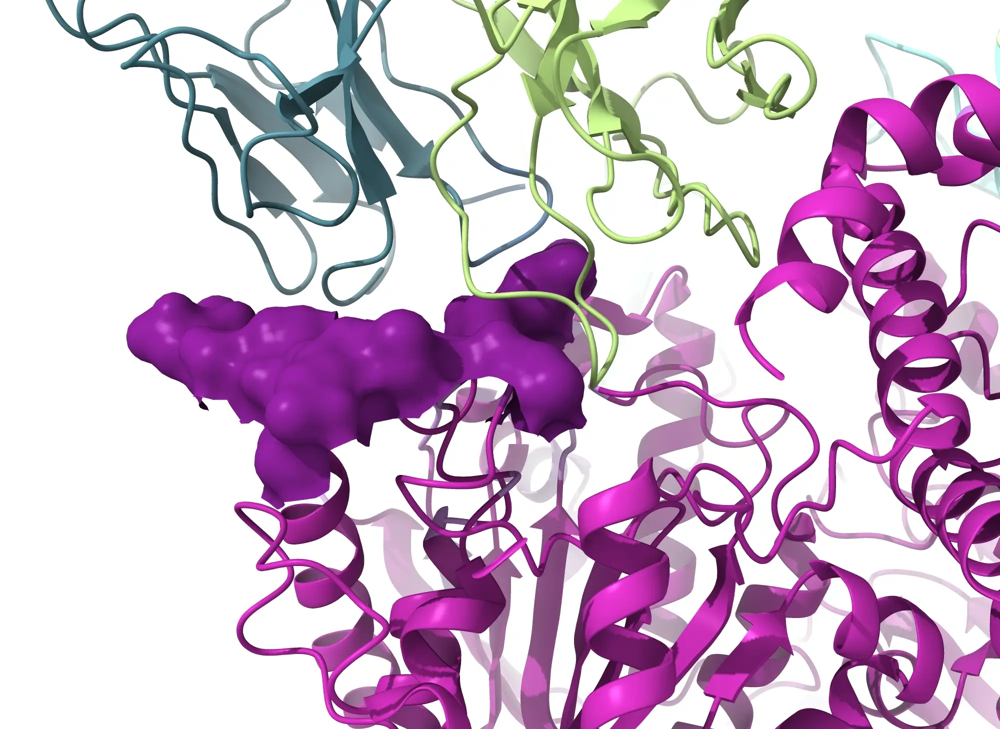
Due to the variety in each sample, our Epitope Mapping bundle begins with an overnight screening in order to assess the suitability of your sample before moving forward.

Have you been able to work with unstructured antigens, such as tau or a-synuclein?

Menu Hex
have-you-been-able-to-work-with-unstructured-antigens-such-as-tau-or-a-synuclein

Yes, NIS has been able to work with unstructured antigens. However in cases with unstructured antigens we can image the Fab and the piece of the antigen that is bound to the Fab and are not able to visualize anything else. It is a complicated process as the peptide is often small, and below the 80 kDa sample size requirement, so we are not always able to give information about binding with respect to the full protein.

Is glycosylation a problem for cryo-EM and antibody complexes? Is it necessary for cryo-EM reconstructions?

Menu Hex
is-glycosylation-a-problem-for-cryo-em-and-antibody-complexes-is-it-necessary-for-cryo-em-reconstructions

No. Glycosylation is not a problem in cryo-EM. Since we are looking at individual particles, and we do not require crystals, even disordered entities like glycans don't really interfere.

The end result is that we will not be able to see them in our micrograph or in our final reconstructions, but they definitely don't affect the final reconstruction at all. So glycosylation is not necessary, since we can work directly with proteins in their native state.

Is there a limit to the number of antibodies bound to a single antigen that can be visualized at the same time?

Menu Hex
is-there-a-limit-to-the-number-of-antibodies-bound-to-a-single-antigen-that-can-be-visualized-at-the-same-time

As long as the antibodies bind to the antigen at different locations, there is no limit to the number of antibodies that can be visualized in one data collection. In fact, having multiple bound antibodies on one antigen can increase the chances of success as it can increase the ordered mass and reduce preferred orientation, making visualizing and aligning particles easier and clearer.

Does NIS have any experience with Epitope Mapping?

Menu Hex
does-nis-have-any-experience-with-epitope-mapping

Yes, in the past two years, NIS has worked on almost 30 epitope mapping projects. In this paper published in Science, you can read more about NIS’s cryo-EM work done to solve an epitope structure. In this project, our scientists were able to use low resolution maps to identify the FAB binding areas and provide expert suggestions for the inactivation mechanism, and higher resolution maps to fully define the epitopes. We were able to overcome preferred orientation by using different grid types and data collection strategies.

Pavel A. Nikitin et al. ,IMM-BCP-01, a patient-derived anti–SARS-CoV-2 antibody cocktail, is active across variants of concern including Omicron BA.1 and BA.2.Sci. Immunol.7,eabl9943(2022).DOI:10.1126/sciimmunol.abl9943

How long will Epitope Mapping with NIS take?

Menu Hex
how-long-will-epitope-mapping-with-nis-take

You can expect to receive a fully refined, 3D map within 2 weeks of sample receipt, assuming your sample meets certain criteria, such as being well-behaved in ice or the epitope not being located on a flexible loop. We understand the challenges associated with samples behaving poorly in ice, and we have developed strategies to overcome these issues. To learn more about our approach to addressing such challenges, we invite you to read our blog post here.

How much does Epitope Mapping with NIS cost?

Menu Hex
how-much-does-epitope-mapping-with-nis-cost

The price to obtain a high-resolution structure, which includes the Initial Starter Pack and additional data collection for mapping, is $35,000. Optional model building is available for $8,800.

Does NIS provide antibody purification or digestion services?

Menu Hex
does-nis-provide-antibody-purification-or-digestion-services

Not at this time. We request that clients provide the system or the complex ready to go on the microscope.

NIS Mountain Hero
NIS Mountain Hero